Call or Text us at:
(905) 877-7171

Digital radiography replaces traditional film with electronic sensors and computer processing to create clear, high-resolution images of teeth, bones, and surrounding tissues. Rather than waiting for chemical development, the sensor captures an image that is transmitted instantly to a computer for review. This shift from analog film to a digital workflow creates a more efficient imaging process and opens up options for enhancing and analyzing images in ways film cannot.
For patients, the experience is quick and straightforward: the sensor is positioned similarly to film, but the resulting image appears on a monitor within seconds. Clinicians can zoom, adjust contrast, and apply measurement tools to examine areas of concern more precisely. These capabilities make digital radiography a versatile diagnostic tool across routine exams, restorative planning, and emergency evaluations.
Although the term “digital x-ray” can sound technical, the practical outcome is simple: faster diagnosis, clearer images, and a record that integrates seamlessly with modern electronic patient files. This combination benefits both patients and clinicians by reducing appointment time and supporting more informed treatment decisions.
One of the most important advantages of digital radiography is the reduction in radiation exposure compared with traditional film techniques. Advances in sensor sensitivity allow clinicians to capture diagnostic-quality images using lower radiation doses. This is especially relevant for patients who require frequent imaging, children, and those with heightened sensitivity concerns.
Comfort improvements are also notable. Modern digital sensors are slim and ergonomically designed, which can make image capture less intrusive than older, stiff film packets. Shorter exposure times and the ability to retake images immediately when positioning needs adjustment further reduce discomfort and anxiety for many patients.
Beyond individual appointments, the environmental footprint of imaging is smaller because digital workflows eliminate the need for hazardous chemicals and film processing. For practices committed to safer, more sustainable care, digital radiography supports both clinical safety and environmental responsibility.
At the center of digital radiography is the sensor — a small electronic device placed in the mouth to detect x-rays and convert them into digital signals. Once captured, the raw image is processed by software that optimizes brightness, contrast, and sharpness, enabling clinicians to identify subtle signs of decay, bone loss, or other issues that might be missed on older film images.
Clinicians can use a range of image-enhancement tools to clarify areas of interest: adjusting contrast to better reveal root structures, magnifying regions to assess margins of restorations, or using measurement features to evaluate bone levels. These tools help dentists make more accurate clinical judgments and communicate findings to patients more clearly during consultations.
After processing, images are stored securely in the patient’s electronic record. Digital storage simplifies appointment workflows by ensuring images are readily available for future comparisons, progress tracking, and coordinated care. Properly managed digital archives also support efficient retrieval when a specialist consultation is needed or when transferring records between offices.
Digital radiography plays a central role across many aspects of dental care. In routine exams, bitewing and periapical images reveal early decay and assess the health of tooth roots. For restorative and prosthetic planning, detailed images help clinicians evaluate existing restorations and determine appropriate treatment margins. In endodontics, high-resolution images are essential for locating canals and verifying the quality of root canal therapy.
Orthodontic assessments, implant planning, and periodontal evaluation also benefit from digital imaging. Clinicians can use sequential images to monitor changes over time — for example, tracking bone levels around teeth or evaluating how an implant integrates with the jawbone. These longitudinal comparisons are easier and more accurate with digital files that can be aligned and reviewed side by side.
In emergency situations, rapid digital imaging is invaluable. When a patient presents with pain, swelling, or trauma, immediate access to clear images allows the dental team to make swift, evidence-based decisions about urgent treatments and follow-up care.
Digital radiography brings advantages in how images are shared and protected. Secure electronic health record systems enable clinicians to share diagnostic images with specialists or referring providers while maintaining patient privacy. Encrypted transmission and access controls help ensure that imaging data is handled according to professional and regulatory standards.
At the office of Mountainview Dental, digital imaging is integrated into the patient care pathway to support transparent communication and collaborative treatment planning. When appropriate, clinicians will review images with patients during their appointment, using on-screen tools to explain findings and the rationale for recommended care. This visual approach helps patients understand their oral health and participate in decisions about treatment.
Maintaining high standards for image quality, storage, and security is a priority. Regular equipment calibration, staff training on image capture and software tools, and adherence to data-protection protocols ensure that digital radiography enhances clinical accuracy without compromising patient confidentiality.
In summary, digital radiography is a safe, efficient, and clinically powerful method for dental imaging. It reduces radiation exposure, improves patient comfort, and provides detailed images that support accurate diagnosis and long-term monitoring. If you would like more information about how digital x-rays are used in our practice or what to expect during an imaging appointment, please contact us for more information.
Digital radiography uses electronic sensors and computer processing to capture dental images instead of photographic film. Images appear on a monitor within seconds, eliminating chemical development and speeding diagnostic workflows. The digital format also allows clinicians to enhance images with tools for contrast, magnification, and measurement.
Compared with film, digital systems often require lower radiation exposures and simplify recordkeeping because files integrate directly into electronic patient charts. Digital images are easier to duplicate and compare over time, which supports more accurate monitoring of disease progression and treatment outcomes. Overall, the shift from analog film to digital sensors improves efficiency and diagnostic flexibility in routine and specialized care.
Digital sensors are more sensitive to x-rays than traditional film, which typically permits diagnostically useful images at lower exposure levels. Clinicians also follow established radiation-safety principles such as using the smallest practical field of view, high-speed imaging settings, and appropriate shielding to protect patients. These measures are particularly important for patients who require periodic imaging or for children with developing tissues.
When imaging is clinically indicated, digital radiography balances diagnostic benefit with safety by minimizing exposure while producing clear images. If you have specific concerns about radiation, the dental team will explain the reasons for imaging and any precautions that will be taken. Clinical judgment guides the frequency and type of imaging to ensure patient safety.
Digital images give clinicians immediate access to high-resolution views of teeth, roots, and supporting bone, which aids in detecting early decay, evaluating restorations, and assessing root health. Software tools allow for zooming, contrast adjustment, and linear measurements that make it easier to identify subtle findings and quantify changes. These enhancements improve diagnostic confidence and help prioritize care based on objective image data.
For treatment planning, digital radiography supports restorative decisions, endodontic procedures, implant placement, and orthodontic assessments by providing precise anatomical information. Sequential images can be compared side by side to monitor healing or disease progression, enabling more effective long-term monitoring. Clear visual documentation also helps clinicians explain conditions and recommended treatments to patients during consultations.
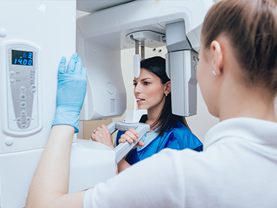
An intraoral digital sensor is positioned in the mouth much like a traditional film packet, and the image is captured with a short exposure while you remain still. The resulting image appears on the clinician's monitor within seconds, allowing immediate review and, if necessary, a quick retake to correct positioning. Sensors are slimmer and more ergonomically shaped than older film packets, which many patients find more comfortable during image capture.
Appointments that include imaging are usually brief, and the dental team will explain each step and answer any questions before taking images. In some cases, extraoral digital imaging such as panoramic radiography may be used for a broader view of the jaws and teeth. The staff will select the most appropriate imaging modality based on the clinical need and the goal of keeping the process as efficient and comfortable as possible.
After capture, raw sensor data is processed by imaging software that optimizes brightness, contrast, and sharpness to highlight diagnostic features. Clinicians can apply tools to magnify regions of interest, adjust contrast to reveal marginal decay or root structure, and use measurement functions to assess bone levels or restoration margins. These manipulations are non-destructive to the original file and are intended to clarify findings rather than alter clinical facts.
Enhanced views help detect early disease and refine treatment decisions, but they are used in combination with a clinical exam and patient history. Images are typically reviewed in real time with patients so the dentist can point out specific concerns and explain treatment rationale. Proper training in image interpretation and software tools is essential to ensure enhancements support accurate clinical judgments.
Digital images are saved directly into secure electronic health records where they can be indexed, archived, and retrieved for future care. Practices maintain access controls, regular backups, and encrypted transmissions to protect patient privacy and ensure data integrity. Staff training, equipment calibration, and software updates are part of routine procedures to maintain consistent image quality and safeguard records.
At the office of Mountainview Dental, imaging files are managed in accordance with professional standards so clinicians can efficiently review past studies and coordinate care when needed. Secure storage also facilitates timely comparison between visits, which helps with monitoring treatment progress and making evidence-based decisions.
Yes. The rapid turnaround of digital images makes them particularly useful in urgent situations such as facial trauma, sudden tooth pain, or suspected infection. Immediate access to clear radiographs allows the dental team to quickly identify fractures, root issues, or areas of bone involvement and to determine the most appropriate urgent interventions. This speed supports practical triage and short-term treatment planning during emergency visits.
Digital imaging also enables clinicians to document acute findings and share images with specialists or emergency providers if referral is required. Having prior digital records available for comparison can be especially helpful in assessing changes that inform follow-up care and definitive treatment planning.
Digital radiography benefits a wide range of patients, including children, individuals who require frequent monitoring, and anyone undergoing complex restorative, endodontic, or implant treatment. The lower exposure levels and quick image capture are advantageous for pediatric care and for patients who may be anxious during dental procedures. Because images are easy to compare over time, patients undergoing long-term treatment plans also gain the most from digital monitoring.
Certain populations, such as pregnant patients, are managed with additional caution and imaging is performed only when clinically necessary and with appropriate shielding. The dental team assesses each patient's needs individually and selects imaging modalities based on diagnostic benefit, safety considerations, and clinical urgency. Open communication about risks and benefits is part of routine patient care.
Digital images can be securely exported and transmitted to specialists, labs, or other dental offices using encrypted electronic transfer protocols that protect patient privacy. Many practices use standardized formats that preserve image quality and metadata, ensuring receiving clinicians have the information needed for coordinated care. Consent and appropriate release procedures are followed when records are shared to comply with privacy regulations.
When referrals are made, images accompany the clinical summary to streamline communication and reduce the need for duplicate imaging. The ability to send high-quality radiographs electronically supports timely consultations and collaborative treatment planning while maintaining strong data-protection practices at the point of transfer.
Maintaining image accuracy requires routine calibration of sensors and x-ray units, regular software updates, and preventive maintenance performed according to manufacturer guidelines. Dental teams also follow quality-assurance protocols such as periodic image audits, staff competency training, and adherence to exposure and positioning standards. These measures help reduce artifacts, minimize repeat exposures, and ensure consistent diagnostic performance.
In addition to equipment care, practices implement strict infection-control procedures for intraoral sensors and establish policies for secure image storage and backup. Ongoing education and adherence to regulatory guidelines combine to keep digital radiography reliable and clinically valuable for everyday patient care.